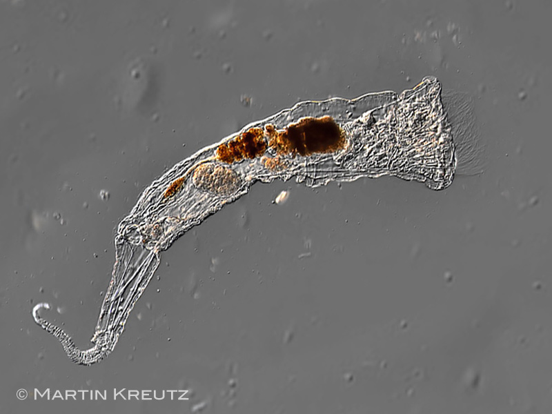
| Collotheca atrochoides, total view. The images of these specimens seem to be the first findings of this species since its discovery by Wierzejski in 1893. In contrast to other Collotheca-species C. atrochoides does not produce a gelatinous sheath, but is free-living in the boundary layer betwen the water column and the anoxic mud of Sphagnum-ponds. |
| |
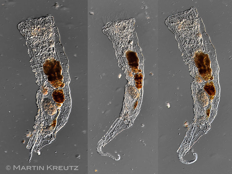 |
| Collotheca atrochoides, specimen with egg; juxtaposition of 3 positions: left: retracted foot; middle: extended corona; right extended foot which does not have an adhesive disk. |
| Here is a video of a living specimen. |
| |
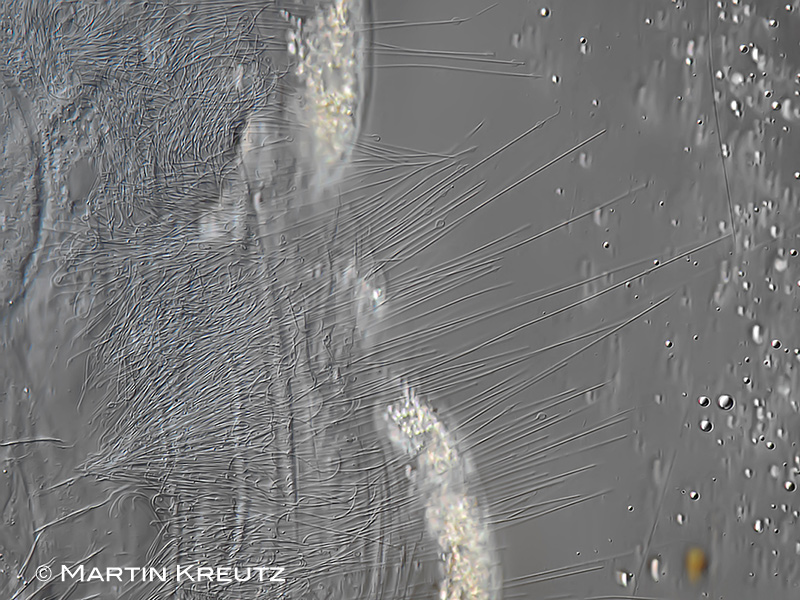 |
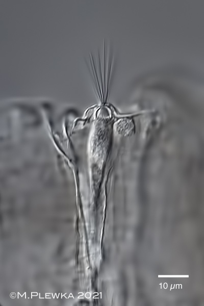
| Collotheca atrochoides, part of the corona. In contrast to Atrochus tentaculatus, which does not have cilia in the corona, the cilia of this species are up to 80 µm long. |
| |
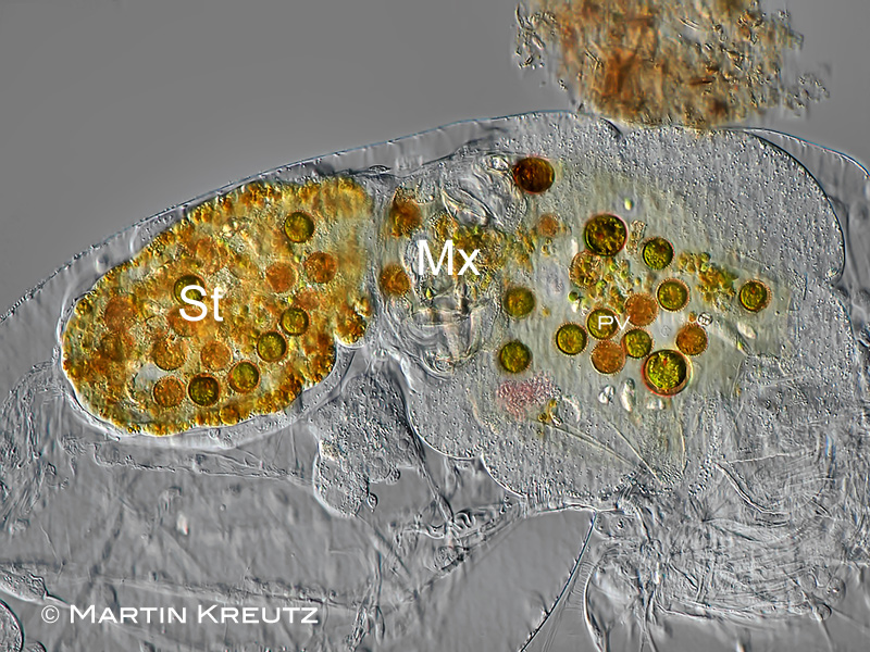 |
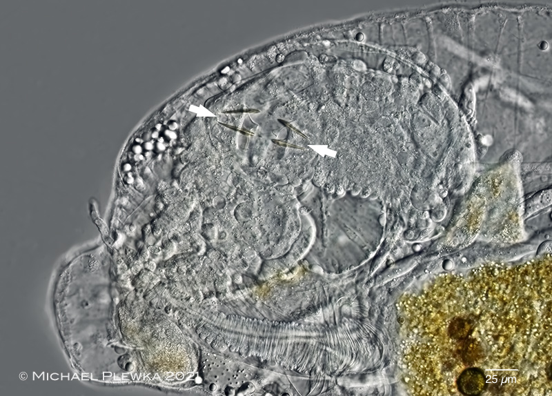
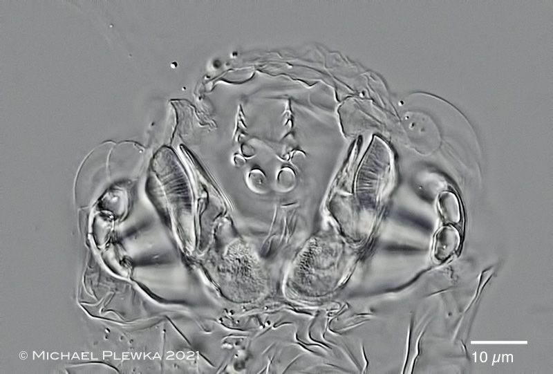
| Collotheca atrochoides, this species could be observed swallowing actively detritus flakes. The diet of this specimen is euglenid flagellates (Trachelomonas sp.), but also Phacus sp./ Euglena sp. or heterotrophic flagellates or rhodobacteria could be observed in the stomach. Mx: Mastax with trophi; PV: proventriculus; St: stomach. |
| |
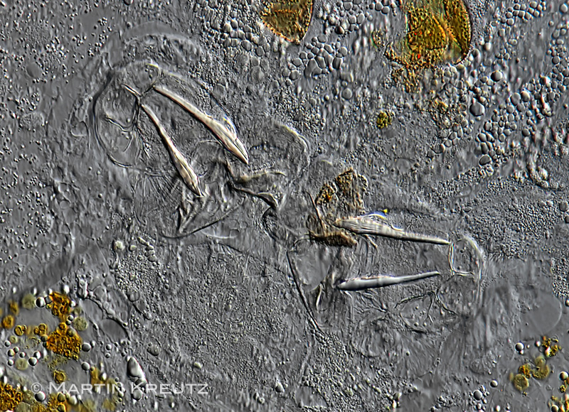 |
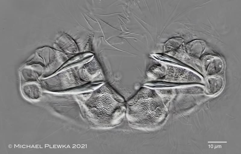
| Collotheca atrochoides, the uncinate trophi are different from the drawing of the trophi of Atrochus tentaculatus by Wierzejski. |
| |
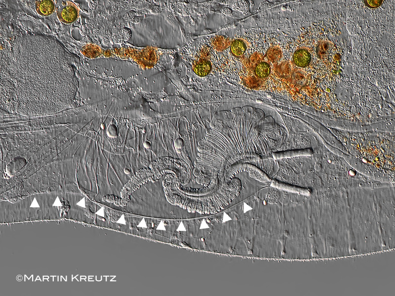 |
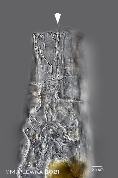
| Collotheca atrochoides, the foot can be retracted completely into the trunk. The triangles mark the border of the "pocket" into which the foot is retracted. |
| |
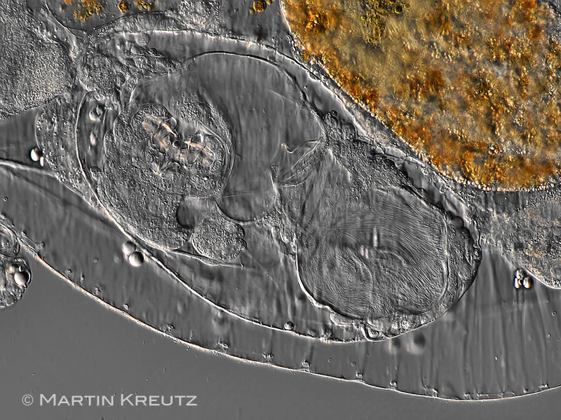 |
| Collotheca atrochoides, embryo; this is one of the few monogonont rotifers which is viviparous. |
| |
| |
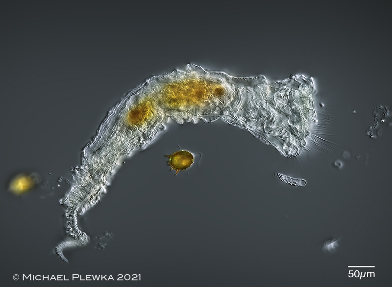 |
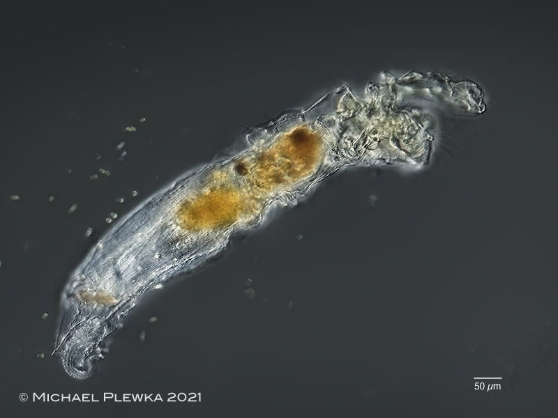
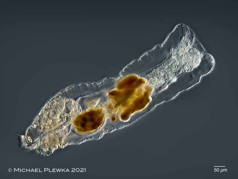
| Collotheca atrochoides, specimen from (3); total view. Two euglenid specimens (species) of the potential prey are surrounding the rotifer (and in focus) in this image: the photosynthetic Trachelomonas armata (left) and the colorless and (osmotrophic) Menoidium cf pellucidum (right). |
| |
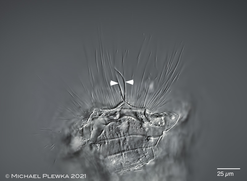 |
| Collotheca atrochoides, frontal part of the ciliated corona. The arrowheads point to two thickened ??sensory?? cilia/ bristles. (3) |
| |
 |
| Collotheca atrochoides, lateral view of specimen from (2); corona partly extended. |
| |
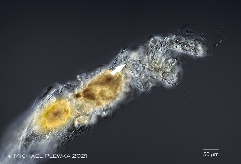 |
| Collotheca atrochoides, lateral view of specimen from (2); corona partly extended. The arrow points to the dorsal antenna. |
| |
  |
| Collotheca atrochoides, left image: dorsal view of specimen with retracted corona. The cilia of the dorsal antenna are visible (arrowhead). Right: crop, showing the dorsal antenna. (2) |
| |
 |
 |
| Collotheca atrochoides, another pregnant female (upper image) and detail of the embryo (lower image). The arrows point to the dents of the unci of the trophi. (3) |
| |
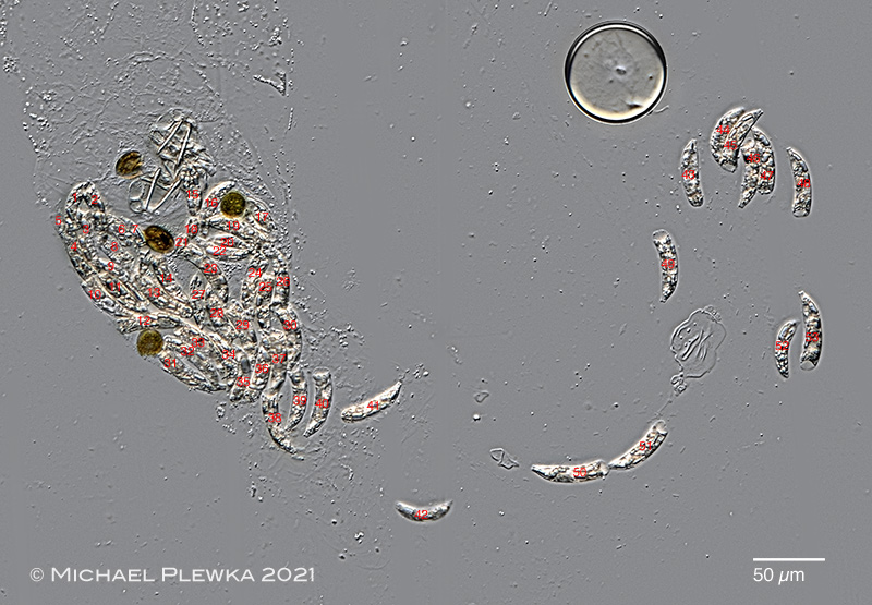 |
| Collotheca atrochoides, content of the proventriculus of a compressed specimen. Like other Collotheca-species the diet of this species seems to be algae that move actively into the proventriculus, in this case euglenid protists which propagate by flagellae. More than 50 Menoidium-cells and 4 Trachelomonas-cells were found in this specimen! See also Collotheca edenta with similar behavior (diet of that species is diatoms which also move actively into the proventriculus). The arrows point to the trophi.(3) |
| |
 |
 |
| Collotheca atrochoides, two images of the uncinate trophi (macerated with SDS). (3) |
| |
| |
| Samples and some images courtesy of Dr. Martin Kreutz, realmicrolife. |
| |
| |
| |
| |
| |
| |
|
|
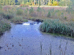 |
| Location(1); (2); (3);: Simmelried near Konstanz, BW, Germany, pond II |
| |
| Habitat (1); (2); (3);: in the boundary layer betwen the water and the anoxic mud (upper 10cm of the mud) (1); (3) |
| |
| Date : 2014 (1); 06.07.2021 (2); 04.09.2021 (3); |
| |
|
|
|
|
|
| |
| |
| |
|
|